Health
Lateral View Of The Eye

The lateral view of the eye is a detailed image of the side of the eye. It shows the eyeball, eyelid, and surrounding structures. The lateral view can be used to assess the health of the eye and to diagnose and treat conditions that affect the side of the eye.
The eye is a complex and fascinating organ, and there is a lot to learn about its various parts and functions. The lateral view of the eye provides a great deal of information about this important part of the body.
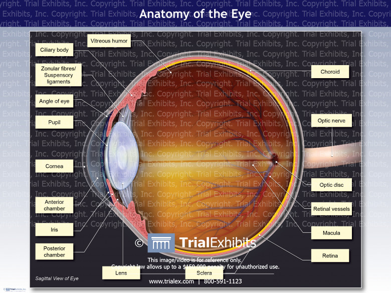
The lateral view of the eye shows the eyeball from the side.
This view can be used to see the different layers of the eye, as well as some of the structures that are located around it. For example, you can see the eyelid, which protects the surface of the eyeball. You can also see the tear duct, which helps to keep the eye moist and healthy.
This view also allows you to see how light enters into the eye. You can see that there is a clear opening at the front of the eyeball, called the pupil. This is where light first enters into the eye.
The pupil gets bigger or smaller depending on how much light is present in your environment.

Credit: quizlet.com
What is the Lateral Part of the Eye?
The lateral part of the eye is the area that is farthest away from the midline of the face. This area includes the side of the eyeball and the surrounding structures, such as the muscles that control eye movement. The lateral part of the eye is important for vision because it contains many of the structures that are responsible for focusing light on the retina.
These structures include the cornea, which is a clear tissue that covers the front of the eye, and the lens, which helps to focus light on the retina.
What is the Side View of Your Eye Called?
The side view of your eye is called the lateral view. The lateral view allows you to see the iris, pupil, and sclera (white part) of the eye. It also lets you see how much of the eyeball is exposed.
How Does the Eye View Images?
The human eye is an amazing organ that is able to view images in a number of different ways. The way that the eye views images is by using a combination of light and dark areas to create an image. This process is known as contrast sensitivity.
The human eye is able to see a wide range of colors, but it is not equally sensitive to all colors. The eye is most sensitive to green and least sensitive to violet. This fact explains why the sky appears blue, because the atmosphere scatters more blue light than other colors.
The human eye can also adjust its focus in order to see objects at different distances. This ability, called accommodation, allows us to see both near and far objects clearly.
So how does the eye actually create an image?
Light enters the eye through the cornea, which bends the light and focuses it onto the retina. The retina is a layer of nerve cells that line the back of the eyeball. These cells are sensitive to light and send electrical signals to the brain when they are stimulated by light waves.
The brain then interprets these electrical signals as images. This process happens so quickly that we are usually unaware of it happening!
Where Do You Perform a Coronal Cut in an Eye Dissection?
A coronal cut is a type of incision made through the front or side of the eye. This approach provides access to the anterior chamber, lens, and vitreous body for surgical procedures such as cataract surgery and vitrectomy. The coronal cut is also used in some types of enucleation (removal of the eye).
The coronal approach begins with an incision made just behind the hairline at the top of the forehead. The incision is then carried down along the side of the eyeball until it reaches the level of the pupil. At this point, another incision is made that extends from just below the pupil to halfway down towards the bottom eyelid.
Finally, a third incision is made across the bottom eyelid to complete the circle around the eye.
After making these incisions, surgeons will typically use scissors or a scalpel to make a small horizontal cut on either side ofthe iris (the colored part ofthe eye). This allows them to peel backthe outer layerof tissue (calledthe conjunctiva) and gain access to underlying structures.
Once inside, surgeons can proceed with their chosen procedure.
While a coronal cut may provide better access for certain types of surgeries, it does have some potential drawbacks. One is that it can cause significant cosmetic damage to the eye area – particularly if visible scars are left behind after healing.
Additionally, this approach carries a riskof injuring important nerves and blood vessels in and aroundthe eye. For these reasons, surgeons must weigh carefully whether or not a coronal approach is right for each individual patient before proceeding with surgery.
Eye anatomy
Lateral View of Eyeball Labeled
The lateral view of the eyeball is a great way to see all the different parts of the eye and how they work together. The cornea, iris, and pupil are all clearly visible in this view. The sclera, or white part of the eye, can also be seen.
This view allows for a good look at how the different parts of the eye work together to allow us to see.
Conclusion
The eye is a complex organ that allows us to see the world around us. The eye has many different parts, all of which work together to allow us to see. The cornea is the clear outer layer of the eye.
It helps to focus light as it enters the eye. The iris is the colored part of the eye. It helps to control how much light enters the eye.
The pupil is the black part of the eye. It controls how much light enters the eye by getting smaller or larger. The lens is a clear structure behind the pupil that helps to focus light on the retina.
The retina is a thin layer at the back of the eye that senses light and sends signals to the brain about what we are seeing.
-
Cloth7 years ago
10 Free Plus Size Clothing Catalogs That You Can Request Online
-
Search Engine Optimization6 years ago
List of 100 High Authority Free Guest Blogging Sites that Bring You Success on The Web
-
Search Engine Optimization6 years ago
The Secret of Link Building Strategies That Works For Every Major Search Engines
-
Blogging2 years ago
How to Start A Blog in 2022 : Step by Step Guide for Beginners
-
Cloth7 years ago
10 Free Junior Clothing Catalogs That You Can Get at Home
-
Email Marketing6 years ago
Methods To Building Your Email List from Blogging
-
Cloth7 years ago
8 Clothing Catalogs for Women That You Can Get for Free
-
Cloth6 years ago
Free Clothing Catalogs That’ll Help You Follow the Latest Fashion Trends


